[ad_1]
Not so long ago, most heart surgeries required open-heart surgery. However, advances in imaging have turned many of these complex surgeries into minimally invasive procedures. Some procedures can now be performed on an outpatient basis, reducing risks and shortening hospital stays. It’s not just invasive procedures that are being innovated. Cardiac care as a whole is evolving, from diagnosis to treatment to follow-up and beyond.
One of the most important advances is the ability to leverage multimodal data. Combining information from a variety of sources, including electrocardiography, echocardiography, computed tomography (CT), magnetic resonance imaging (MRI), and positron emission tomography (PET), all enable AI. , clinicians will be able to see a more holistic view. The patient’s condition. A deeper understanding of each patient’s unique situation allows physicians to individualize treatment plans, increasing the likelihood of success.
“AI-enabled devices can provide better, faster images, lower radiation doses, and improved workflows,” explains Eigil Samset, general manager of cardiology solutions at GE HealthCare.
Because each modality targets a different aspect of heart disease, insights can be extracted by combining data across different modalities and even across time, allowing clinicians to see trends and correlations in new ways. Masu. When physicians can easily access and visualize all relevant data, they can better determine the safest and most effective treatment options for an individual.
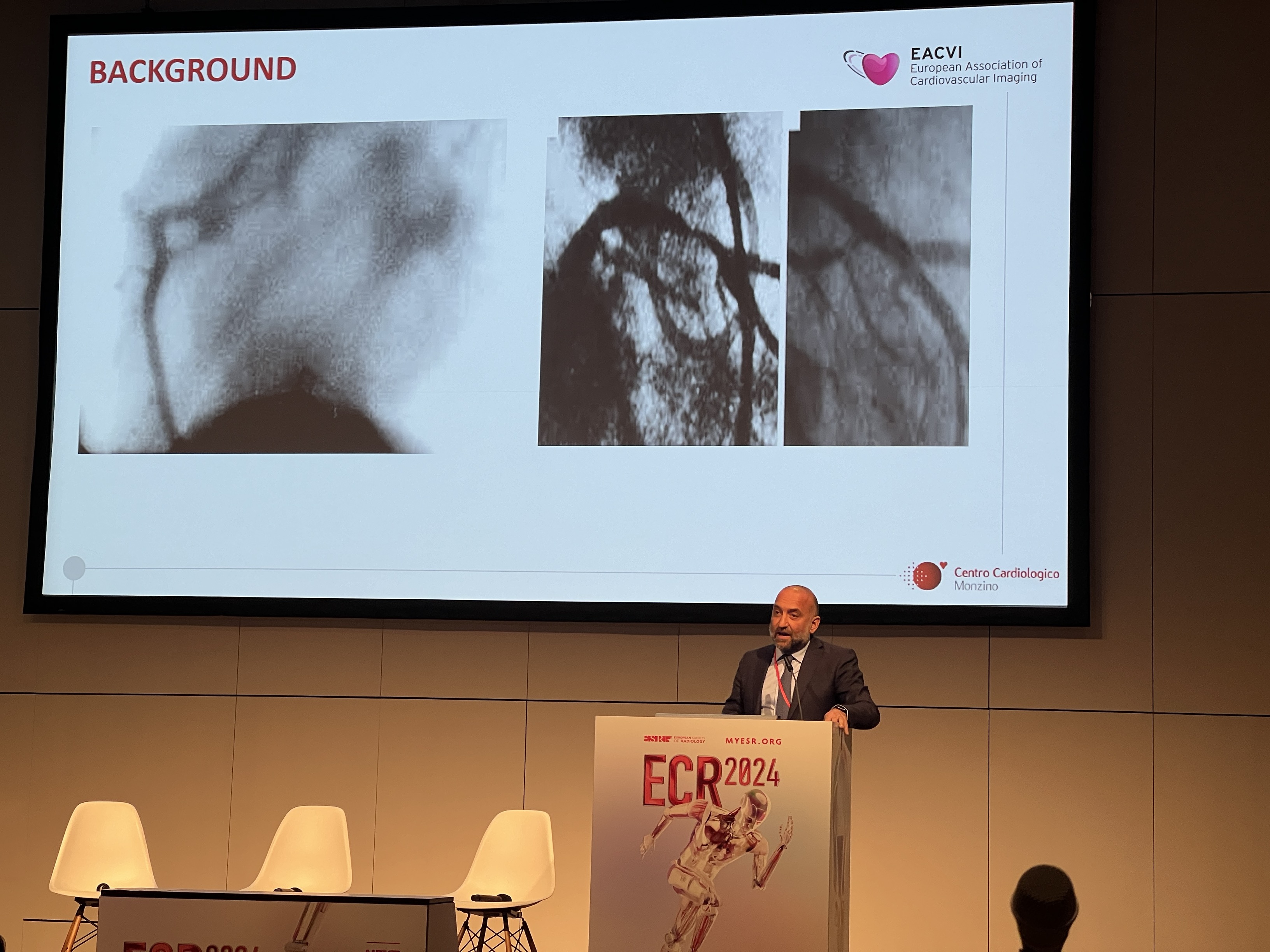
Professor Gianluca Pontone, director of perioperative cardiology and cardiovascular imaging at Italy’s Centro Cardiologico Monzino, has previously investigated invasive angiography for possible coronary artery disease. He explains that there was a prejudice against treating patients with stents even if they had coronary artery disease. An arterial bypass graft (CABG) would have been a better option.
Professor Gianluca Pontone
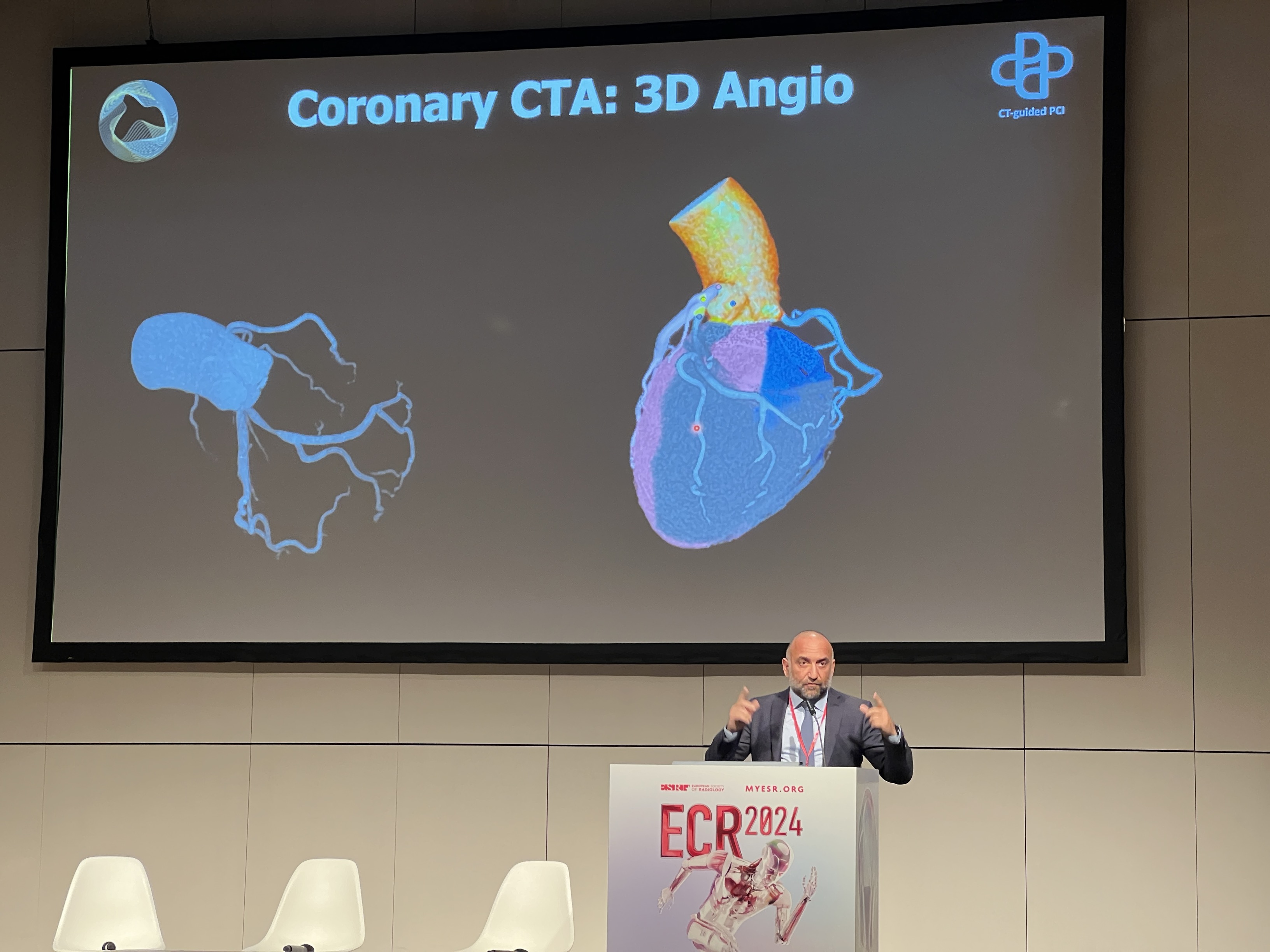
“Thanks to the combination of computed tomography (CT) equipment and perfusion assessment, we now know in advance which patients should be treated with drug therapy, who will benefit from PCI, and who will be more appropriate for CABG. “We can now analyze a patient’s anatomy and calculate a score that is used in clinical practice,” says Ponton.
The ability to visualize exactly what is happening in a non-invasive setting relieves clinicians from time pressure during surgery. The entire team, including imagers, interventionists, and surgeons, can discuss each patient’s condition and take the most appropriate steps to ensure the best outcome with the least amount of risk.
Multimodality imaging combined with 3D software also helps surgeons visualize the heart’s complex anatomy, allowing them to plan approaches before surgery and guide tools during surgery. . “Cardiac computed tomography is becoming a very important tool in planning procedures, for example to ensure that catheters are properly sized to avoid rupture during treatment,” Pontone said. says. .
Patients are not the only ones who benefit. “Non-invasive imaging takes the guesswork out of the equation and provides great peace of mind when you can calculate the likely outcome of a strategy,” he continues. “And this is very important because there are often scenarios in the medical world where the black and white is not clear and the best option is not clear.” But these AI-powered tools , to help you get oriented. ”
Regular multimodal imaging can track the progression of heart disease and the patient’s condition, allowing cardiologists to make adjustments as needed and avoid complications such as leaks or blood clot formation after valve replacement surgery. You can specify.
“We are building a digital fabric that connects everything across the patient care pathway,” Samsett said. “Rather than just collecting images and data, we can actually give insights back to caregivers and use that to understand how patients are doing and improve access to care. ”
Multimodal imaging is also beginning to be developed as a predictive tool. Currently, heart disease is diagnosed and treated when a patient shows symptoms, but clinicians are using multimodal technology to predict which patients need treatment before the classic signs of heart disease appear. I’m starting to use image processing. Early intervention can extend the lifespan of patients and give them a better quality of life. Treating patients before they show signs of illness also has the potential to reduce overall treatment costs.
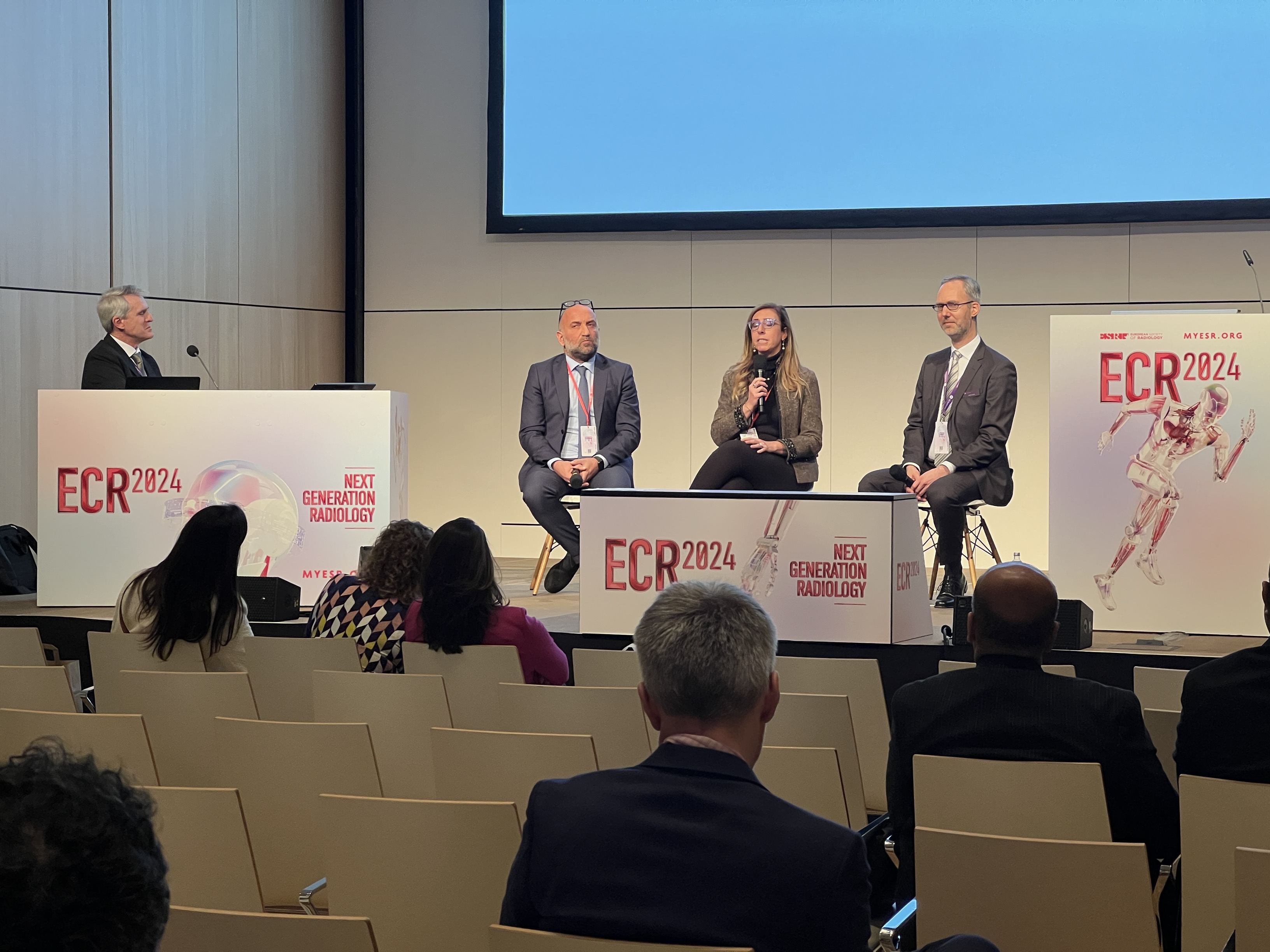
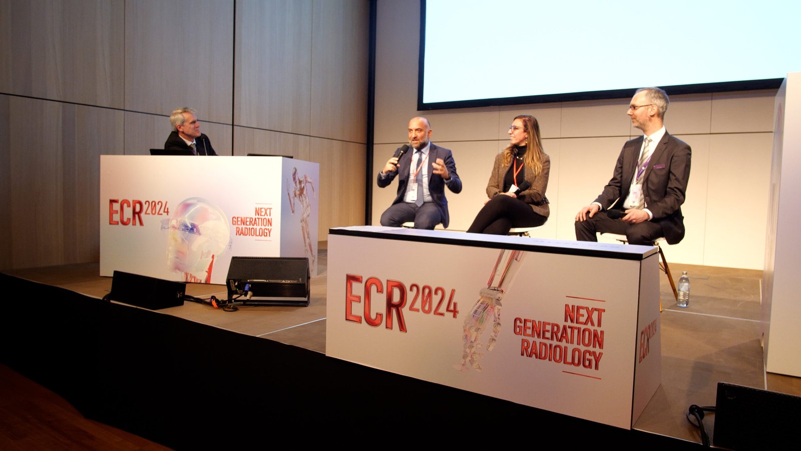
From left to right: Professor Gianluca Pontone, Professor Chiara Bucciarelli Ducci, and Martin Janic from GE HealthCare.
“There is emerging evidence that there are patients with preclinical heart disease who are asymptomatic, may benefit from treatment and intervention, and who can be identified with imaging tests,” Pontone said. “We may be able to identify new subsets of asymptomatic patients, in which case treatment may become reasonable.”
Powerful advances in AI-powered multimodal imaging are making these tools more powerful and versatile, helping clinicians better understand how heart disease is impacting individual patients. This revolutionized cardiac medicine. This allows us to tailor treatment plans to each patient’s specific needs and track progress, ultimately resulting in better treatment outcomes and healthier patients.
Â
![]()
[ad_2]
Source link
