
[ad_1]
Author: Phoebe Ingraham Renda
From the Oculus gaming system to Apple’s Vision Pro, Pitt students are well-versed in virtual reality (VR) and augmented reality (AR) technology. But for the first time in fall 2023, 115 pharmacy students worked on research using the technology during a cardiovascular disease pharmacotherapy course.
“We teach students how to use drugs to treat different types of heart disease, but before they can do that effectively, they need a solid refresher on the basics of heart anatomy. ” says course instructor and professor James “Jim” Koons. Department of Pharmaceutical Therapeutics, Faculty of Pharmacy. This course is part of the Doctor of Pharmacy program, a 6-year entry-level professional program.
This course has used human patient simulators from Pitt’s Peter M. Winter Institute for Simulation Education and Research since the mid-2000s to provide students with an interactive learning experience, but the anatomy lessons are primarily remained lecture-based.
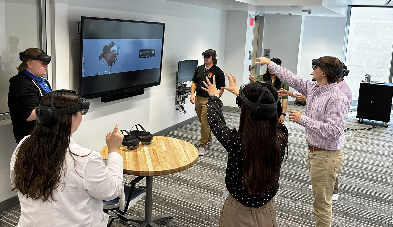
Pharmacy students take a cardiac anatomy class in 3D using HoloLens 2 AR headsets in Alan McGee Scaife Hall. Photo credit: Lawrence Kobrinski.
With a little help from Pitt Health Sciences Information Technology (HSIT), last year’s students moved away from PowerPoint slides and used an Anatmage Table, which resembles a dining table-sized iPad, and a HoloLens 2 AR headset to explore anatomy. I immersed myself in the lesson.
“It’s a million miles away from what Jim used to do,” says instructional designer Lawrence “Larry” Kobrinski, who has helped sponsor the course since 2013. This was unimaginable just a few years ago. ”
Combining AR technology with Anatomage modules for various medical conditions and diagnoses dramatically enriched student learning. You can now stand in front of the 3D heart and explore beyond the heart chambers. Students can manipulate the heart’s arteries and conduction system, examine the anatomy of healthy and unhealthy hearts, and monitor drugs to see where they are working in the heart.
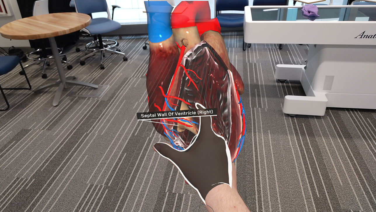
Using HoloLens technology, Pitt School of Pharmacy students physically (and virtually) manipulate 3D cardiac anatomy. Photo by Alexi Zukas
Coons and Kobulinsky are extending their research using AR and patient simulator training to virtual worlds. They are actively creating realistic VR clinical cases with the help of SimX, the leading VR simulation training platform for doctors, military personnel, and emergency medical personnel.
“When you enter a virtual environment, you pick up an electronic medical record (EMR) tablet and scroll through pages, talk on the phone, view patient information, insert IVs, and enter orders from the EMR tablet. ,” says Kobrinski. .
In VR, students interact with critically ill patients, nurses, and doctors within an infectious disease environment. As students interact with the virtual patient, they can see lab changes in the EMR as well as real-time changes such as a drop in blood pressure or other signs that the patient has become unstable. Patients may even develop new complaints that should be considered.
“This technology will transform the way students learn and transform the interdisciplinary learning environment so that students can practice across disciplines,” said Amy Seibert, dean of the School of Pharmacy. “This adds realism to learning. You need to react to patient responses in real time so you can adjust drug therapy or discuss new approaches with your multidisciplinary team.”
Patient details extend to genetics and incorporate training in pharmacogenomics. In this training, students look for specific genes that predict response to drugs and influence treatment recommendations.
“Pharmacogenomics is talked about a lot in cardiology,” Koons says. He said susceptibility to anticoagulants such as warfarin and other drugs that patients take after having a stent (a mesh coil that helps keep an artery open) inserted may vary based on genetics. explains.
Coons and Kobulinsky hope to receive a beta test copy of the VR case this spring and have it ready for use in the fall. For fall classes, Koons and Kobrinski will also include evaluations to show that teaching with these technologies increases student learning and retention. “This is definitely going to be part of the future of pharmacy education,” Seibert says.
Kobrinski, who grew up with iPads and new technology, points out that today’s students have different learning expectations than students who came here 15 to 20 years ago. Digitally native students easily become immersed and enthused in helping simulated patients, recalling information they have read, heard, and seen in other courses. “This allows them to integrate everything into this digitally connected world that they’re used to,” Seibert said.
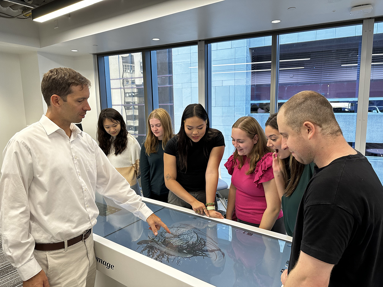
Jim Coons (left), professor in the Department of Pharmacy and Therapeutics in the School of Pharmacy, uses an anatomy table to teach students an anatomy lesson. Photo credit: Lawrence Kobrinski.
This fall, the class will be taught in a brand new AR/VR lab located on the mezzanine floor of Alan McGee Scaife Hall and will be available to all Health Sciences Schools. The new space will feature a variety of AR/VR headsets (including HoloLens 2, Meta Quest 3, and Vive Pro 2), an Anatomage Table, and a large video recording studio for live streaming and recording lectures.
“We’re looking at how pharmacies used this technology last year when planning new spaces,” said Jane Alexander, assistant director of educational technology at HSIT. “We hope to generate interest from other groups in the health sciences and showcase what technology can do and how it can benefit learning.”
Seibert, Koons and Kobrinski hope this technology will be implemented in other courses in the School of Pharmacy and inspire other courses in the School of Health Sciences to transform historical didactic lectures into interactive 3D learning experiences. I’m looking forward to it.
“This is Dr. Shekhar’s vision for the health sciences, and we are excited to be a part of it and partner with each of the health science schools,” Seibert said.
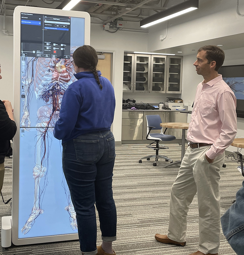
Koons watches students explore life-sized heart and circulatory anatomy on an anatomy table. Photo credit: Lawrence Kobrinski.
[ad_2]
Source link
